
Upper cervical thermography
Explore ChiroTherm’s upper cervical thermography information and professional resources.
Revolutionizing Chiropractic Diagnosis
Advanced Infrared Thermography for Upper Cervical Spine & Whole-Body Health
For the upper cervical chiropractor, Infrared spectrum technology has provided our doctors with an enhanced ability to locate and identify subluxation and its systemic effects of the entire body. Not only is it an invaluable tool in the evaluation of subluxation, it can also provide objective evidence of other health conditions such as stroke, cardiovascular disease, inflammation, neuropathy, and cancer.
Beyond Paraspinal Analysis: A New Frontier in Subluxation Evaluation
What You Need to Know
Comprehensive Benefits of Infrared Thermography
Infrared thermography provides a non-invasive view of surface temperature patterns. These findings may support clinical assessment and progress monitoring when interpreted alongside a patient history and physical examination.
Utilization

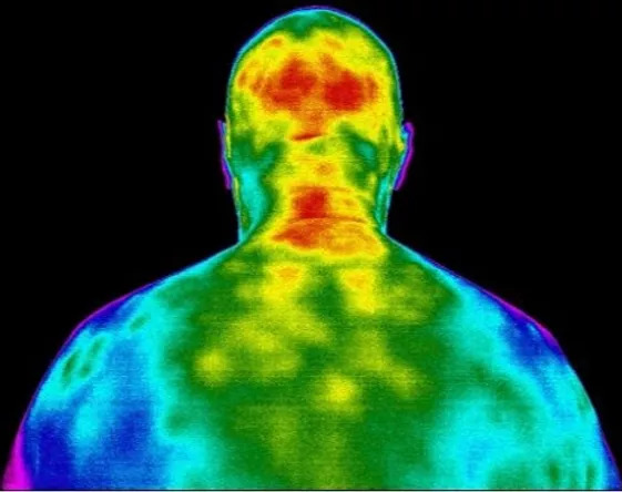
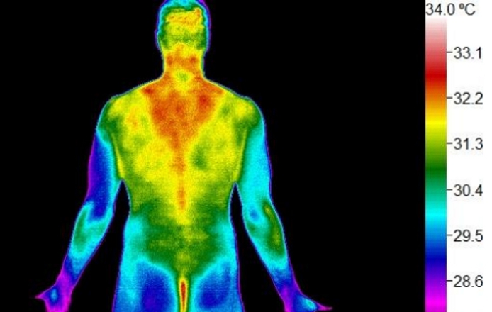
Infrared thermography measures patterns of heat emitted from the body’s surface. Clinicians may use these patterns alongside the patient history and physical examination to identify areas that warrant closer evaluation. Potential uses include:
- Measure neurovascular function
- Identify spinal inflammation
- Locate tissue damage
- Monitor recovery and healing
Applications

- Neurological Conditions
- Spinal Subluxation
- Vertigo and Headaches
- Spinal Damage
- Pinched Nerve
- Numbness and Tingling
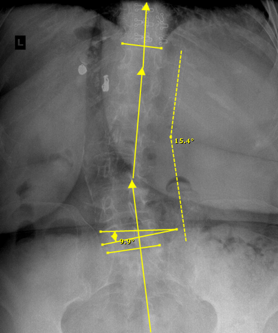
- Childhood and Adult Scoliosis
- Sciatica and Low Back Pain
Safety

The best part about infrared thermography is that it does not emit anything into the body, it only detects the heat that is coming out.
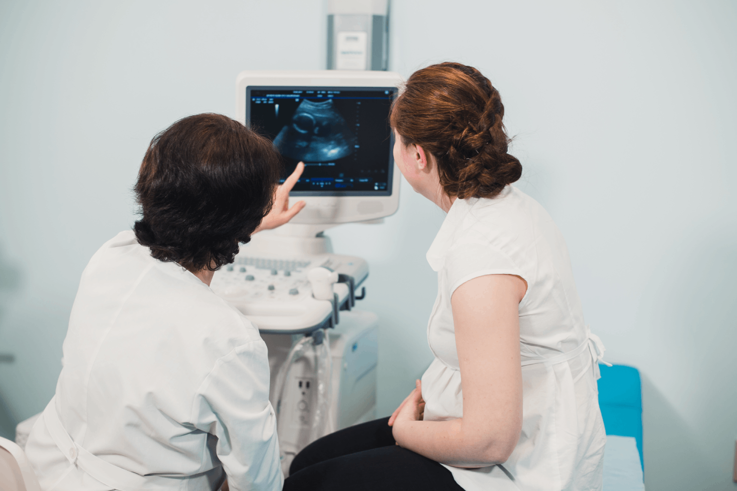
- Safe for Pregnancy
- Pediatric and Newborn Safe
- Painless and Quick
- Non-Invasive
- No Radiation
Guided Integration
Integrating Thermography Is Simple
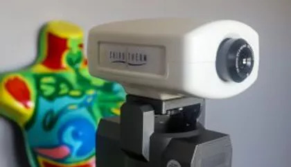
- Choose a camera for your practice. Our experts will help you decide .
- Design your Thermography Room with help from our staff
- Receive certification through PACT
- Evaluate your patient images or send in for interpretation
- Create a customized program for your patients
- Use thermography daily or to re-evaluate upon completion of program.
- Can be used live during treatment as a video
Focused Approach
Upper Cervical & Chiropractic Applications
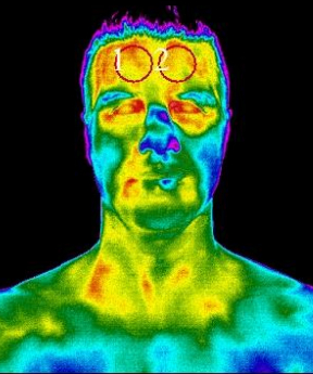
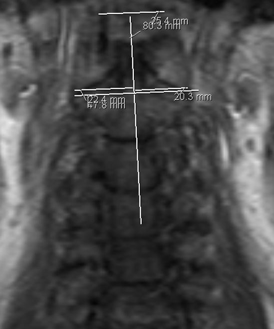
Frontal
- Cerebrovascular Screening
- Facial Nerve Evaluation
- Oral and Orbital Evaluation
- Sinus Visualization
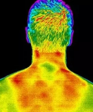
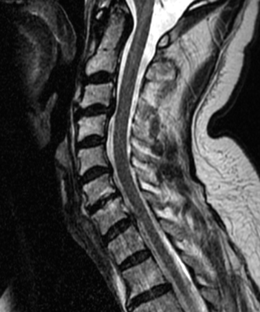
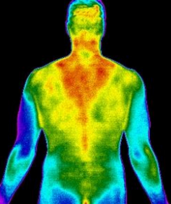
Posterior
- Nerve Impingement
- Paraspinal Symmetry
- Myotomal Evaluation
- Dermatomal Evaluation
- Inflammation
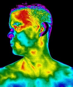
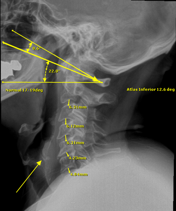
Lateral
- Symmetry Evaluation
- Anterior Atlantal Tissue
- Lymphatic System
- Auditory Canal Evaluation
- Temporal Artery Function
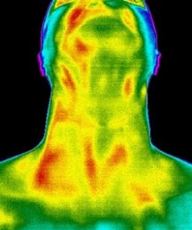
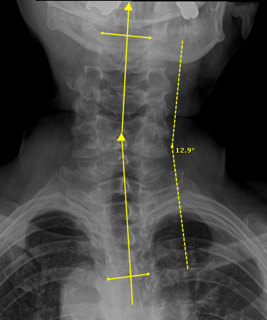
Anterior
- Carotid Artery Evaluation
- Thyroid Evaluation
- Lymph Evaluation
- Scalene Symmetry
Full Spine
- Systemic Effect
- Radiculopathy
- Adaptive Stress
- Global Inflammation
Additional Applications
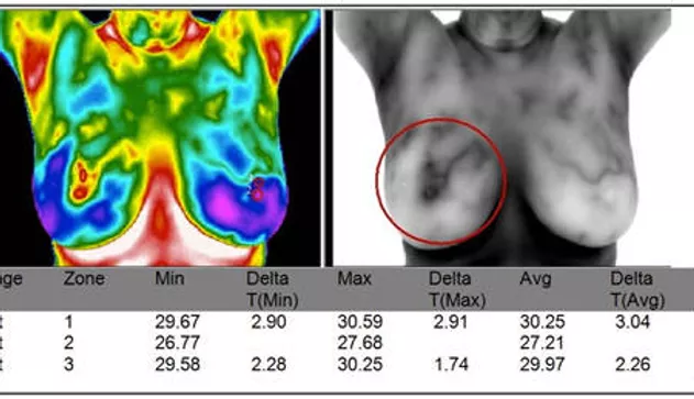
Breast Thermography
Breast thermography is a non-invasive, radiation-free, and painless method for screening breast health. It uses state-of-the-art medical infrared technology to detect and assess heat patterns in breast tissue. By establishing an individual thermal baseline through comparative exams, thermography helps monitor subtle physiological changes over time—providing valuable insights into early signs of inflammation or potential abnormal activity.
With breast thermography, we analyze heat patterns in the breast that may signal underlying pathology. Tumor growth often leads to increased vascular activity, which generates heat that can be detected by advanced infrared cameras. Thermography is especially useful for assessing fibrocystic breasts and provides a safe, non-invasive way to monitor overall breast health.
Thermography should always be used as a tool in addition to other diagnostic testing
Thyroid Pathology

Thermal imaging may show surface-temperature differences in the neck, but it does not diagnose thyroid disease or replace laboratory testing, ultrasound, or medical evaluation. Any findings should be interpreted in clinical context.
Cerebrovascular Patterns

Thermal imaging can show differences in facial and forehead surface temperature. These findings are nonspecific and should not be used alone to diagnose impaired circulation, carotid blockage, or stroke. Concerning symptoms require prompt medical evaluation.
Neuro-Muscular

Impingement upon a nerve can alter normal blood flow, leading to observable physiological changes. In some cases, affected dermatomes or myotomes may be visible through a reduction in surface heat. These areas present as hypothermic patterns on thermal imaging, providing insight into potential nerve compression or dysfunction.
Postural Assessment
Postural stress places abnormal strain on joints, particularly on the side of deviation. Thermal imaging can identify the resulting inflammation or ischemia in these cases. The plantar surfaces of the feet are highly sensitive and typically exhibit increased heat on the side of postural stress, increased weight bearing, or compensatory gait abnormalities.